


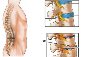
Lumbar osteochondrosis is a chronic disease that develops as a result of degenerative-dystrophic process in the intervertebral discs. The disease is widespread and affects most people between the ages of 25 and 40.
According tostatistics, every second adult experiences back pain at least once, and in 95% of cases it is caused by osteochondrosis of the spine.
Patients with severe lumbar osteochondrosis, persistent pain, and other manifestations recognize a temporary disability. If their condition does not improve within four months, the issue of setting up a disability group will be resolved.
Lumbar osteochondrosis is a serious medical and social problem, as the disease mainly affects people of most working age and in addition, in the absence of treatment, it can lead to the formation of a herniated disc.
Causes and risk factors
Factors contributing to the development of lumbar osteochondrosis are:
- spinal structure abnormalities;
- Lumbarization - a congenital pathology of the spine, characterized by the separation of the first spine from the spine and its transformation into the sixth (additional) lumbar;
- Sacralization is a congenital pathology in which the fifth lumbar spine is combined with the sacral; Asymmetric layout of joint spaces of the intervertebral joints;
- Abnormal narrowing of the spinal canal;
- reflects spondiogenic pain (somatic and muscular);
- obesity;
- sedentary lifestyle; Prolonged exposure to
- vibration;
- systemic physical stress;
- smoking.
Prolonged compression of nerve roots, which over time irritate certain organs in the abdomen, causes their function to deteriorate.
Instability of the spinal segment is accompanied by reactive changes in the bodies of the surrounding spine, intervertebral joints, and accompanying spondyloarthritis. For example, significant contraction of muscles in the face of physical exertion causes the vertebral bodies to move and the nerve roots to deceive with the development of radicular syndrome.
Another cause of lumbar osteochondrosis pain and neurological symptoms may be osteophytes - bone growths on processes and vertebral bodies that cause radicular syndrome or compression myelopathy (spinal cord compression).
Forms of the disease
Depending on which structures are involved in the pathological process, lumbar osteochondrosis is clinically manifested by the following syndromes:
- reflex- lumbodinia, lumboshalgia, lumbago; Develops against the background of reflex overload of the back muscles;
- Compression (spinal, vascular, radicular)- Compression (compression) of the spinal cord, blood vessels, or nerve roots causes them to develop. Examples are lumbosacral sciatica, sciatica.
Symptoms of lumbar osteochondrosis
Symptoms in lumbar osteochondrosis are determined by which structures are involved in the pathological process.
Lumbago occurs under the influence of hypothermia or physical stress, sometimes for no apparent reason. The pain appears suddenly and is of a shooting nature. It is strengthened by wheezing, coughing, body twitching, exercise, sitting, standing, walking. Pain sensations in the supine position are significantly attenuated. Sensitivity and reflexes are maintained, the range of motion of the lumbar spine is reduced.
Observe on palpation:
- pain in the lumbar region;
- sphincter muscle spasm; Flatness of the lumbar lordosis, often combined with scoliosis.
Nerve root tension syndrome in Lumbago is negative. When lifting a straight leg, patients notice an increase in pain in the lumbar region and not on their outer side in the lower extremity.
Often, lumbar osteochondrosis causes recurrent pain attacks that become more intense and prolong each time.
The clinical picture in Lumbodinia is similar to that of Lumbago, but the increase in pain intensity occurs within a few days.
During lumboizalgia, patients complain of pain in the lumbar region that radiates to one or both lower extremities. The pain spreads to the buttocks and the back of the thigh and never reaches the legs.
Lumboshalgia is characterized by vasomotor disorders:
- Changes in skin temperature and color of the lower extremities;
- heat or cold;
- Circulatory disorders.
The development of lumbar compression syndromes is clinically manifested by the following symptoms:
- dermatomal hypalgesia;
- shooting pains; weakening or complete loss of
- deep reflexes;
- Peripheral paresis.
Compression syndromes are aggravated by pain in the trunk, coughing and coughing.
Diagnostics
Diagnosis of lumbar osteochondrosis is made on the basis of clinical picture of the disease, laboratory and instrumental research methods.
Blood tests for lumbar osteochondrosis:
- Decreased calcium concentration;
- increased ESR;
- increased alkaline phosphatase levels.
X-ray examination of the spine is of great importance in the diagnosis of lumbar osteochondrosis.
Prolonged compression of nerve roots, which over time irritate certain organs in the abdomen, causes their function to deteriorate.
X-ray signs that confirm the diagnosis are:
- Change the configuration of the affected segment;
- Pseudospondylillolysis (movement of adjacent vertebral bodies);
- deformation of closing plates;
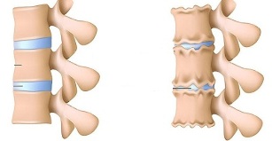
- flatness of the intervertebral disc;
- Uneven height of the intervertebral disc (spacer symptom) associated with asymmetric muscle tone.
Also used in the diagnosis of lumbar osteochondrosis, if indicated,
- Myelography, computed tomography or magnetic resonance imaging - necessary for the development of permanent symptoms, neurological deficits;
- Scintigraphy (study of phosphorus accumulation in the bone system, labeled tech-99) - is performed if there is a suspicion of a tumor or an infectious process, spinal cord injury.
Differential diagnosis of lumbar osteochondrosis is made with the following diseases:
- spondylolisthesis;
- dyshormonal spondylopathy;
- ankylosing spondylitis (ankylosing spondylitis);
- Infectious processes (disc inflammation, spinal osteomyelitis);
- neoplastic processes (primary tumor of the spine or its metastatic lesion);
- rheumatoid arthritis;
- Osteoarthritis deformity of the hip joint;
- reflects pain (diseases of internal organs and large blood vessels).
Treatment of lumbar osteochondrosis
In lumbar osteochondrosis, the following treatment tactics are usually followed:
- Bed rest for 2-3 days;
- traction of the affected segment of the spine;
- Strengthening the spinal muscles and abdominal muscles (creating a so-called muscle corset);
- Influence on pathological myofacial and myotonic processes.
Lumbago occurs under the influence of hypothermia or physical stress, sometimes for no apparent reason.
In most cases, conservative treatment of lumbar osteochondrosis is performed, including the following:
- Infiltrative muscle anesthesia with a solution of local anesthetics;
- taking nonsteroidal anti-inflammatory drugs; taking
- desensitizers;
- Vitamin therapy; taking
- tranquilizers and antidepressants;
- Manual therapy, massage;
- Physiotherapy exercises;
- acupuncture;
- Post-isometric rest.
Absolute indications for surgical treatment of lumbar osteochondrosis are:
- Acute or subacute compression of the spinal cord;
- development of cauda equina syndrome, characterized by pelvic organ dysfunction, sensory and motor disorders.
Therapeutic exercises for lumbar osteochondrosis
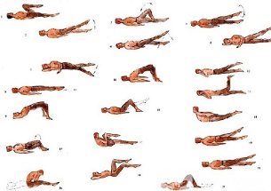
Physical therapy plays an important role in the complex treatment of lumbar osteochondrosis. Regular exercise allows you to regulate the muscle tone of the paravertebral muscles, improve metabolic processes in the tissues affected by the pathological process, and in addition create a well-developed muscle corset that can help the correct position of the spine to relieve unnecessary static loads.
For gymnastics with lumbar osteochondrosis, which gives the greatest effect, you should follow the following principles:
- regularity of lessons;
- a gradual increase in the intensity of physical activity;
- Avoid overloading during the lesson.
Physiotherapy should be conducted under the guidance of an experienced instructor who selects the exercises that are most effective for a particular patient and monitors the correctness of their implementation.
According tostatistics, every second adult experiences back pain at least once, and in 95% of cases it is caused by osteochondrosis of the spine.
In addition to lessons with the instructor, you should conduct a daily set of morning exercises that include special exercises for lumbar osteochondrosis.
- Relax and compress the abdominal muscles.Standing position, legs shoulder-width apart, hands down on body. Smooth breathing, relaxation of the muscles of the anterior abdominal wall. Pull the stomach as much as possible while exhaling, tense the abdominal muscles. Exercise should be repeated until light fatigue appears.
- Head movements with spinal flexion.Starting position is on your knees, with your arms outstretched on the floor, with your back straight. Slowly lift your head and lean on your back. Hold this position for a few seconds, then smoothly return to the starting position. Repeat at least 10-12 times.
- "Pendulum".Starting position Lying on your back, arms along the body, legs should be bent at right angles to the knee and hip joints. Rotate your legs to the right and left, in pendulum-like movements, to try to reach the floor. In this case, the shoulder blades cannot fall off the floor.
- boat.Initial position: lie on your stomach with your arms outstretched. Destroy the upper body and legs off the floor, back bent. Hold this position for 5-6 seconds and slowly return to the starting position. Run 10 times.
Potential Outcomes and Complications
The main complications of lumbar osteochondrosis are:
- Interstitial hernia formation;
- vegetative-vascular dystonia;
- spondylolysis, spondylolisthesis;
- osteophytosis;
- spondyloarthritis;
- Spinal canal stenosis, which has caused spinal cord injury and can lead to permanent disability and reduced quality of life.
forecast
Pain syndrome in lumbar osteochondrosis occurs in the form of remissions and exacerbations. Lumbago lasts 10-15 days, after which the patient's condition improves, the pain subsides. Favorable outcome can be avoided by associated secondary diseases. Often, lumbar osteochondrosis causes recurrences of pain attacks that become more intense and prolong each time.
Physical therapy plays an important role in the complex treatment of lumbar osteochondrosis.
Patients with severe lumbar osteochondrosis, persistent pain, and other manifestations recognize a temporary disability. If their condition does not improve within four months, the issue of setting up a disability group will be resolved.
Prevention
Prevention of osteochondrosis of the spine includes the following measures:
- Cigarette smoking
- normalize body weight;
- Improving general physical condition, active lifestyle; Avoid provocative conditions (weight lifting, sudden movements, twisting, bending).